Electrophoresis: Separation of Charged Molecules
Electrophoresis is a technique used to separate charged particles (such as DNA, RNA, proteins) based on their size and charge by applying an electric field. Molecules migrate through a gel or liquid medium at different speeds: smaller/faster or more highly charged molecules move farther. It is essential in molecular biology, forensic science, clinical diagnostics, and biochemistry.
Principle of Electrophoresis
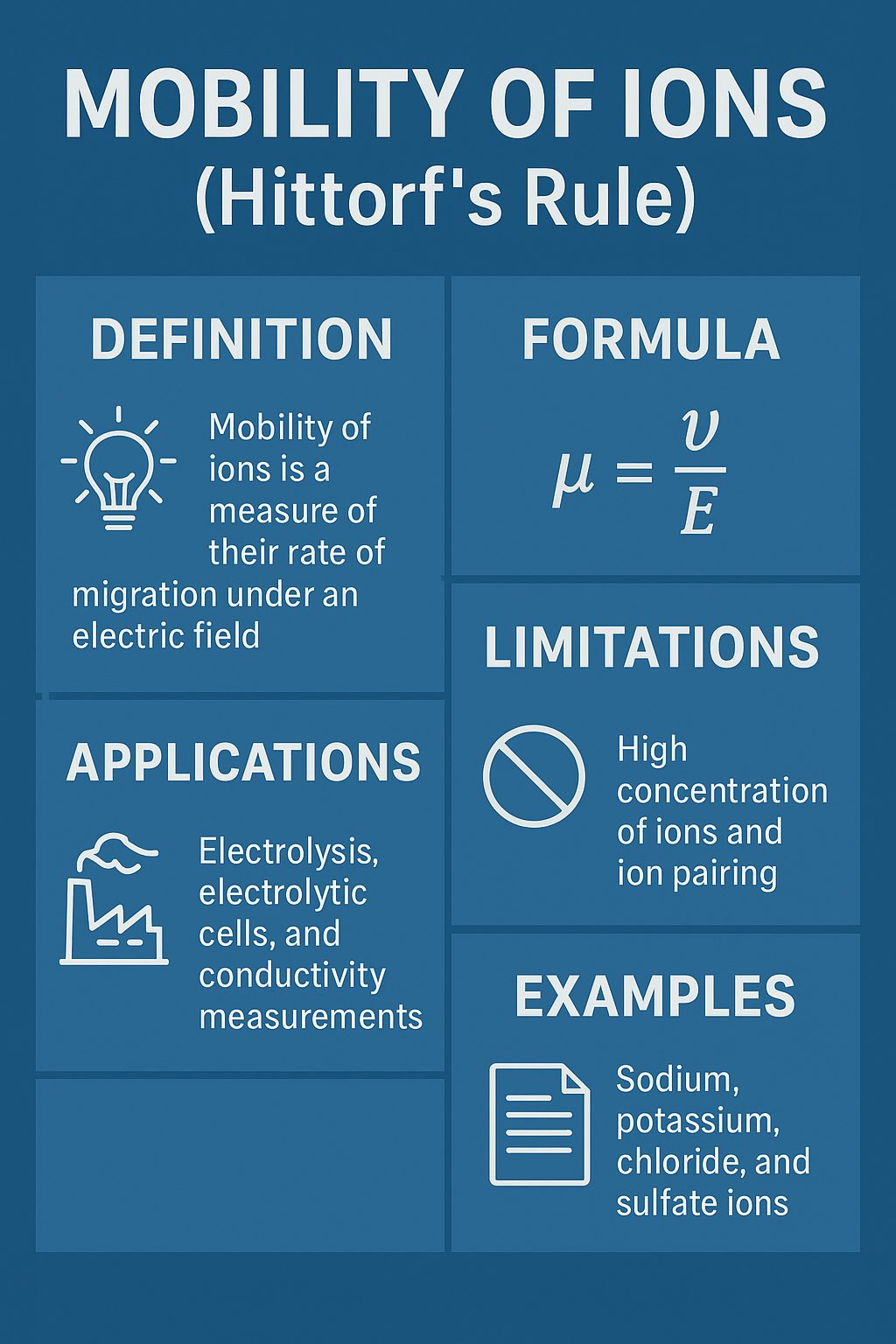
Charged molecules placed in an electric field experience a force proportional to their net charge. They migrate toward the oppositely charged electrode. The matrix (gel, capillary) provides resistance that separates molecules by size: smaller molecules move faster through the pores. The electrophoretic mobility (μ) depends on charge, size, shape, and medium viscosity.
Types of Electrophoresis
🧬 Gel Electrophoresis (Agarose/PAGE)
Separates DNA fragments, RNA, or proteins by size using an agarose or polyacrylamide gel. Visualised with stains (EtBr, Coomassie).
🧪 Capillary Electrophoresis (CE)
High-resolution separation in narrow capillaries. Fast, automated, used in DNA sequencing and pharmaceutical analysis.
⚡ SDS-PAGE (Proteins)
SDS denatures proteins and imparts uniform negative charge; separation by molecular weight only.
🧬 Pulsed‑Field Gel Electrophoresis (PFGE)
Alternating electric fields separate very large DNA molecules (e.g., whole chromosomes).
🧫 Isoelectric Focusing (IEF)
Separates proteins by their isoelectric point (pI) using a pH gradient.
🔬 Zonal Electrophoresis
Uses a density gradient to stabilise separated zones; used in clinical labs.
🎮 Continuous Particle Simulation: Electrophoretic Separation
🧪 Real‑time Electrophoresis Simulation
Adjust voltage (electric field) and select sample type. Charged particles (or molecules) move from the top (negative) toward the bottom (positive electrode). Different molecules move at different speeds based on their size-to-charge ratio. Smaller/faster molecules travel farther in the same time.
💡 In the simulation, each band represents a group of molecules (DNA fragments or proteins). Higher voltage increases migration speed. Different colours indicate different molecular sizes. Reset restarts the run.
Applications of Electrophoresis
- DNA fingerprinting / forensics: Matching crime scene samples.
- Diagnostics: Detection of genetic mutations, sickle cell anaemia, HIV.
- Protein analysis: Purity check, molecular weight determination.
- Pharmaceutical QC: Purity of biopharmaceuticals (insulin, antibodies).
- Environmental microbiology: Identification of microbial communities.
Factors Affecting Electrophoretic Mobility
- Net charge: Higher charge → greater mobility.
- Size & shape: Smaller, globular proteins move faster.
- Electric field strength: Higher voltage → faster migration.
- Buffer pH: Determines ionisation state of molecules.
- Gel concentration: Higher agarose % slows larger DNA fragments.
📝 Electrophoresis – Quiz
1. What is the driving force for separation in electrophoresis?
2. Which technique is used to separate DNA fragments by size?
3. In SDS-PAGE, proteins are separated based on:
4. Which dye is commonly used to visualise DNA in agarose gels?
5. Increasing the voltage in electrophoresis typically:
Comprehensive notes covering all types, applications, and troubleshooting.
Download Complete Notes Below
Proudly Powered By


Leave a Comment